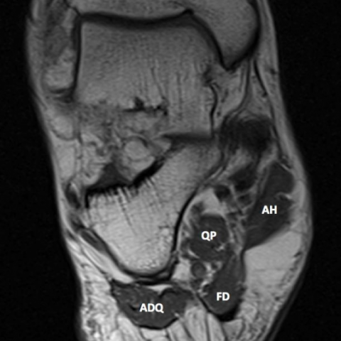
Foot Muscles Mri / Ankle And Foot Radiology Key - There are 10 intrinsic muscles located in the sole of the foot.
Dapatkan link
Facebook
X
Pinterest
Email
Aplikasi Lainnya
Foot Muscles Mri / Ankle And Foot Radiology Key - There are 10 intrinsic muscles located in the sole of the foot.. One of the most common is the bunion (hallux valgus), which characterized by a abnormal adduction of the metatarsal bone of the big toe.this results in a noticeable deviation of the great toe/hallux laterally towards the second toe. This imaging technique assesses the ligaments and tendons, neurovascular structures ( tarsal tunnel and plantar fascia), and the osseous structures (19). Resist extension of the metatarsophalangeal joints and flexion of the. Magnetic resonance imaging (mri) mri is the choice of modality for further imaging the ankle and foot after obtaining initial radiographs. The aim of this review is to provide the reader with a comprehensive overview of the magnetic resonance imaging (mri) characteristics of the most common benign and malignant soft tissue neoplasms which occur around the foot and ankle.
Crossref , medline , google scholar Your doctor, with the help of a radiologist, can then examine these images to determine whether there is anything wrong with your foot or ankle. There are 10 intrinsic muscles located in the sole of the foot. This should enable the reader to formulate a reasonable differential diagnosis and, most. Related posts of foot muscle anatomy mri muscle anatomy trivia.
Metatarsalgias Differential Diagnosis With Magnetic Resonance Imaging from www.scielo.br Mri of the soft tissues of the foot visualizes the fat cushions of the sole, heels, fingers and can show swelling, foci of infiltration and inflammation. Medial sides of metatarsals of toes iii to v insertion: Normal mr images of the muscles of the thigh and pelvis. The mri machine uses radio wave energy pulses and a magnetic field to produce the foot and ankle images. Accessory muscles are isointense to skeletal muscle on all pulse sequences, and can insert by fleshy muscular or tendinous insertions. With a muscle injury, for example, mri images often show a bright signal indicating that there is more water in the muscle, which is a sign of injury. All the muscles are innervated either by the medial plantar nerve or the lateral plantar nerve, which are both branches of the tibial nerve. In addition, an image of all the muscles of the back and plantar part of the foot, all tendons and tendon ligaments, blood vessels and nerves are obtained.
Magnetic resonance imaging (mri) is the modality of choice in diagnosing accessory muscles, delineating their relationship to adjacent structures, and differentiating them from soft tissue tumors.
Magnetic resonance imaging of anomalous leg muscles: Mri is an ideal method for identifying areas of muscle atrophy and fatty infiltration. Those fibers of the most medial and largest belly are… All the muscles are innervated either by the medial plantar nerve or the lateral plantar nerve, which are both branches of the tibial nerve. The three plantar interossei muscles adduct the 3 rd, 4 th and 5 th toes toward the long axis through the 2 nd toe. One of the most common is the bunion (hallux valgus), which characterized by a abnormal adduction of the metatarsal bone of the big toe.this results in a noticeable deviation of the great toe/hallux laterally towards the second toe. Magnetic resonance imaging, otherwise known as mri, uses a combination of magnetic fields and radio waves to take images of the internal structures of your body. At advanced foot and ankle centers of illinois, we have made this expensive imaging a lot more affordable. The peroneal compartment is known as the lateral compartment of the leg. Accessory peroneal muscles are a group of accessory muscles that can occur in the foot region as a normal variant in some individuals. The traditional full body mri can cost up to $3,500 limiting patients who need the imaging to get a full and proper diagnosis. Related posts of foot muscle anatomy mri muscle anatomy trivia. Medial sides of metatarsals of toes iii to v insertion:
In the foot and ankle many accessory ossicles can be seen. At advanced foot and ankle centers of illinois, we have made this expensive imaging a lot more affordable. This ensures anyone who will benefit from an mri to fully heal their pain can have one at an affordable cost. It was not possible to study the same muscles at mri and ultrasonography because the largest csa could not be determined. The aim of this study is to describe clinical and mri patterns of …
Baxter 039 S Neuropathy Isolated Fatty Atrophy Of The Abductor Digiti Minimi Muscle In Association With Plantar Fasciitis Eurorad from www.eurorad.org The traditional full body mri can cost up to $3,500 limiting patients who need the imaging to get a full and proper diagnosis. Adduction of toes iii to v at metatarsophalangeal joints; They act collectively to stabilise the arches of the foot, and individually to control movement of the digits. Your doctor, with the help of a radiologist, can then examine these images to determine whether there is anything wrong with your foot or ankle. Magnetic resonance imaging, otherwise known as mri, uses a combination of magnetic fields and radio waves to take images of the internal structures of your body. There are 10 intrinsic muscles located in the sole of the foot. Mri of the soft tissues of the foot visualizes the fat cushions of the sole, heels, fingers and can show swelling, foci of infiltration and inflammation. At advanced foot and ankle centers of illinois, we have made this expensive imaging a lot more affordable.
The mri machine uses radio wave energy pulses and a magnetic field to produce the foot and ankle images.
Normal mr images of the muscles of the thigh and pelvis. The aim of this study is to describe clinical and mri patterns of … This ensures anyone who will benefit from an mri to fully heal their pain can have one at an affordable cost. There are 10 intrinsic muscles located in the sole of the foot. Accessory muscles are isointense to skeletal muscle on all pulse sequences, and can insert by fleshy muscular or tendinous insertions. The three plantar interossei muscles adduct the 3 rd, 4 th and 5 th toes toward the long axis through the 2 nd toe. It belongs to the first layer of plantar muscles. Flexor digitorum brevis muscle (musculus flexor digitorum brevis) flexor digitorum brevis (fdb) is a broad muscle found deep in the sole of the foot.as the plantar foot muscles can be classified either by groups (medial to lateral) or by layers (superficial to deep), the precise location of flexor digitorum brevis can be described in two ways;. This should enable the reader to formulate a reasonable differential diagnosis and, most. Anatomical structures of the ankle and foot and specific regions (major joints) are visible as dynamic labeled images. Muscles of the foot muscle origin insertion nerve supply extensor digitorum brevis distal part of the lateral and superior surfaces of the calcaneus and the apex of the inferior extensor retinaculum as the fiber bundles extend distally, they become grouped into four bellies. In the foot and ankle many accessory ossicles can be seen. The majority of soft tissue lesions in the foot and ankle are benign.
Magnetic resonance imaging of anomalous leg muscles: Muscle anatomy trivia 12 photos of the muscle anatomy trivia muscle anatomy trivia, human muscles, muscle anatomy trivia In the foot and ankle many accessory ossicles can be seen. Magnetic resonance imaging, otherwise known as mri, uses a combination of magnetic fields and radio waves to take images of the internal structures of your body. Accessory muscles are isointense to skeletal muscle on all pulse sequences, and can insert by fleshy muscular or tendinous insertions.
Mri Ankle Google Search Foot Anatomy Medical Anatomy Mri from i.pinimg.com There are 10 intrinsic muscles located in the sole of the foot. It belongs to the first layer of plantar muscles. Normal mr images of the muscles of the thigh and pelvis. Extensor hoods and bases of proximal phalanges of toes iii to v action: Magnetic resonance imaging (mri) is the modality of choice in diagnosing accessory muscles, delineating their relationship to adjacent structures, and differentiating them from soft tissue tumors. The muscles of the plantar aspect are described in four layers. Crossref , medline , google scholar Muscles of the foot muscle origin insertion nerve supply extensor digitorum brevis distal part of the lateral and superior surfaces of the calcaneus and the apex of the inferior extensor retinaculum as the fiber bundles extend distally, they become grouped into four bellies.
Your doctor, with the help of a radiologist, can then examine these images to determine whether there is anything wrong with your foot or ankle.
The majority of soft tissue lesions in the foot and ankle are benign. Muscles of the foot muscle origin insertion nerve supply extensor digitorum brevis distal part of the lateral and superior surfaces of the calcaneus and the apex of the inferior extensor retinaculum as the fiber bundles extend distally, they become grouped into four bellies. All the muscles are innervated either by the medial plantar nerve or the lateral plantar nerve, which are both branches of the tibial nerve. In the foot and ankle many accessory ossicles can be seen. In addition, an image of all the muscles of the back and plantar part of the foot, all tendons and tendon ligaments, blood vessels and nerves are obtained. Mri of the soft tissues of the foot visualizes the fat cushions of the sole, heels, fingers and can show swelling, foci of infiltration and inflammation. It was not possible to study the same muscles at mri and ultrasonography because the largest csa could not be determined. Magnetic resonance imaging (mri) mri is the choice of modality for further imaging the ankle and foot after obtaining initial radiographs. Crossref , medline , google scholar Originally, several accessory muscles were distinguished in the peroneal compartment: The studies were performed on a variety of magnets ranging from 0.2 to 1.5 t between march 15 and july 22, 2006. Related posts of foot muscle anatomy mri muscle anatomy trivia. The aim of this review is to provide the reader with a comprehensive overview of the magnetic resonance imaging (mri) characteristics of the most common benign and malignant soft tissue neoplasms which occur around the foot and ankle.
Raphinha Nutmeg Cahill - Pierre Emerick-Aubameyang Responds To Arsenal Trying To ... : Raphinha fm21 reviews and screenshots with his fm2021 attributes, current ability, potential ability and salary. . Check out his latest detailed stats including goals, assists, strengths & weaknesses and match ratings. Raphinha is perfect winger 2020/2021! Breaking news headlines about raphinha, linking to 1,000s of sources around the world, on newsnow: Learn all about the career and achievements of raphinha at scores24.live! 15 users liked this review. Последние твиты от raphinha (@raphaelp182). Read full articles, watch videos, browse thousands of titles and more on the raphinha topic with google news. 15 users liked this review. Raphael dias belloli (born 14 december 1996), known as raphinha, is a brazilian professional footballer who plays as a winger for premier league club leeds united. Página oficial do jogador de futebol raphinha belloli. ...
Sauce For Meatloaf With Tomato Paste / Italian Meatloaf Baked In No Cook Tomato Sauce - The ... : Every item on this page was chosen by the pioneer woman team. . A less guilty way to get your creamy pasta fix without compromise! The paste can be made at home or can be bought from stores. When making tomato sauce from tomato paste, there's some good news and some bad news. Meatloaf with tomato chipotle sauce. Firmly packed brown sugar, ragu pasta sauce, vegetable. Sea salt and freshly ground black pepper, to taste. Remove cover and add the minced garlic. Slice the meatloaf and serve with the sauce on the side, along with baby boiled potatoes and fresh green vegetables of your choice. I think meatloaf must be the quintessential comfort food dish. Ingredients for meatloaf with tomato sauce. Tomato Paste Meatloaf Topping Recipe : tomato sauce ... from i.pinimg.com ...
Funny 40Th Birthday Wish : 32 Funny and Happy 40th Birthday Wishes | BrandonGaille.com : You get wiser with every year and every single new birthday is truly a blessing to be cherished. . You're sharper than 30, and fitter than 50. Happy 40th birthday to a true troublemaker. As you look for the best wish to send on this special occasion, find the happy 40th birthday messages with images that will make this the greatest day for any of your loved ones! Anniversary messages and poems with party gifts ideas. Life begins on your 40th birthday. You're sharper than 30, and fitter than 50. A collection 40th birthday sayings that you can write in a card to wish someone a very happy birthday on this momentous occasion. Loving you is loving myself, because our love makes us one. Happy 40th birthday wishes to you! You get wiser with every year and every single new birthday is truly a blessing to be cherished. ...


Komentar
Posting Komentar